
What dermoscopy diagnoses
Differentiates benign moles from melanoma, evaluates basal cell and squamous cell skin cancers, diagnoses fungal scalp infections, alopecias, scabies, lichen planus, psoriasis, vasculitis and many pigmentation disorders.
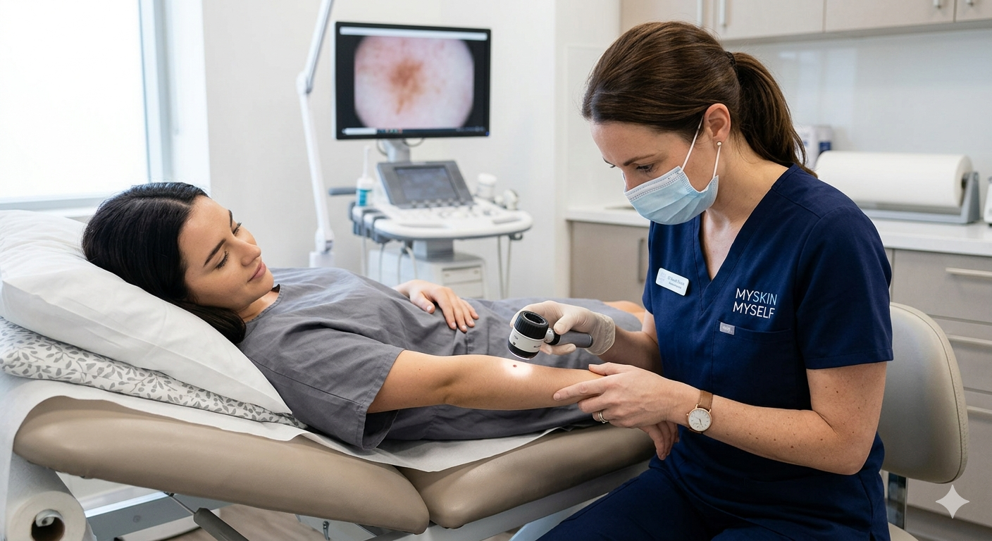
How it is performed
A polarised-light dermoscope is placed on the skin lesion at 10×–20× magnification. Images are captured and stored against your medical record for follow-up comparison — essential for monitoring moles over years.
Why it reduces unnecessary biopsies
Many lesions look concerning to the eye but show benign patterns under dermoscopy — saving needles, scars and worry. Conversely, truly suspicious lesions are caught early.
Dermoscopy in hair and nails
Trichoscopy (scalp dermoscopy) and onychoscopy (nail dermoscopy) help diagnose hair loss subtypes and nail tumours respectively — services included in our Gachibowli dermatology workup.